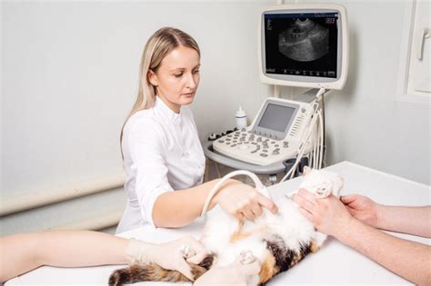
Veterinary ultrasound technology has become an essential diagnostic tool in veterinary medicine, allowing practitioners to non-invasively examine the internal structures of animals. As a veterinary professional, mastering ultrasound technology can significantly enhance your diagnostic capabilities and improve patient outcomes. Here are five tips to help you achieve mastery in veterinary ultrasound technology:
Tip 1: Understand the Fundamentals of Ultrasound Physics

To effectively use veterinary ultrasound technology, it is crucial to understand the fundamental principles of ultrasound physics. This includes knowledge of frequency, wavelength, resolution, and artifacts. A solid grasp of these concepts will enable you to optimize image quality, identify potential errors, and accurately interpret ultrasound images.
Key Concepts in Ultrasound Physics
- Frequency: The number of oscillations per second, measured in Hertz (Hz).
- Wavelength: The distance between consecutive oscillations, measured in millimeters (mm).
- Resolution: The ability to distinguish between two objects, measured in millimeters (mm).
- Artifacts: Errors or distortions in the ultrasound image, caused by factors such as reverberation, refraction, or attenuation.
Tip 2: Familiarize Yourself with Ultrasound Equipment and Settings

Veterinary ultrasound equipment is designed to produce high-quality images of various body structures. To achieve optimal results, it is essential to understand the different settings and features of your ultrasound machine. Familiarize yourself with the following:
Key Features of Ultrasound Equipment
- Transducer selection: Choose the correct transducer for the desired application, considering factors such as frequency, footprint, and materials.
- Gain settings: Adjust the gain to optimize image quality, taking into account the type of tissue being examined and the presence of artifacts.
- Depth settings: Set the correct depth to ensure that the region of interest is visible, while minimizing unnecessary tissue scanning.
- Focus settings: Adjust the focus to optimize image resolution, considering the depth and type of tissue being examined.
Tip 3: Develop Your Scanning Skills through Practice and Training

Mastering veterinary ultrasound technology requires hands-on practice and training. Develop your scanning skills by:
Scanning Tips and Tricks
- Start with simple examinations, such as abdominal scans, and gradually move to more complex procedures.
- Practice on a variety of patients, including dogs, cats, and exotic animals.
- Use ultrasound phantoms or models to develop your scanning skills in a controlled environment.
- Attend workshops, seminars, and online courses to stay updated on the latest techniques and technologies.
Tip 4: Stay Up-to-Date with the Latest Advances in Veterinary Ultrasound

The field of veterinary ultrasound is rapidly evolving, with new technologies and techniques being developed continuously. Stay current with the latest advances by:
Staying Current with the Latest Advances
- Attending conferences, seminars, and workshops focused on veterinary ultrasound.
- Subscribing to veterinary ultrasound journals and publications.
- Participating in online forums and discussion groups dedicated to veterinary ultrasound.
- Joining professional organizations, such as the International Veterinary Ultrasound Society (IVUS).
Tip 5: Integrate Ultrasound into Your Diagnostic Workflow

To maximize the benefits of veterinary ultrasound technology, it is essential to integrate it into your diagnostic workflow. This includes:
Integrating Ultrasound into Your Workflow
- Using ultrasound as a primary diagnostic tool, rather than a secondary or confirmatory modality.
- Combining ultrasound with other diagnostic modalities, such as radiography and computed tomography (CT).
- Developing a standardized ultrasound protocol for common applications, such as abdominal scanning.
- Staying organized and efficient, using tools such as ultrasound report templates and image archiving software.
By following these five tips, you can master veterinary ultrasound technology and enhance your diagnostic capabilities, ultimately improving patient outcomes and the overall quality of care.
Gallery of Veterinary Ultrasound Images


What is the difference between B-mode and M-mode ultrasound?
+B-mode ultrasound produces a two-dimensional image of the body structure being examined, while M-mode ultrasound produces a one-dimensional image of the motion of the structure over time.
How do I choose the correct transducer for a particular examination?
+Choose a transducer with the correct frequency, footprint, and materials for the desired application, considering factors such as tissue depth, resolution, and artifacts.
What is the role of gain settings in ultrasound imaging?
+Gain settings control the amplitude of the ultrasound signal, allowing for optimization of image quality and reduction of artifacts.
