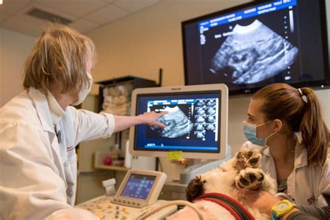
Veterinary care has come a long way in recent years, with advancements in medical technology playing a significant role in improving the diagnosis and treatment of animals. One of the most important diagnostic tools in veterinary medicine is the X-ray, also known as radiography. X-rays are a type of diagnostic imaging that uses X-rays to produce images of the internal structures of an animal's body. In this article, we will explore the world of X-ray technology for animals, its applications, benefits, and how it works.
How X-ray Technology Works for Animals
X-ray technology for animals is similar to that used in human medicine. The process involves exposing the animal to a controlled amount of X-ray radiation, which passes through the body and creates an image on a digital sensor or film. The resulting image, known as a radiograph, shows the internal structures of the animal's body, including bones, organs, and tissues.
The X-ray machine used in veterinary medicine is designed specifically for animals and is typically smaller and more compact than those used in human medicine. The machine is equipped with a digital sensor or film that captures the X-ray image, which is then displayed on a computer screen for the veterinarian to interpret.
Applications of X-ray Technology in Veterinary Medicine
X-ray technology has a wide range of applications in veterinary medicine, including:
- Orthopedic diagnosis: X-rays are commonly used to diagnose bone fractures, joint disorders, and other orthopedic conditions in animals.
- Dental diagnosis: X-rays are used to diagnose dental problems, such as tooth abscesses, fractures, and periodontal disease.
- Thoracic diagnosis: X-rays are used to diagnose respiratory problems, such as pneumonia, pleurisy, and lung cancer.
- Abdominal diagnosis: X-rays are used to diagnose gastrointestinal problems, such as intestinal blockages, tumors, and foreign bodies.
Benefits of X-ray Technology for Animals
X-ray technology has numerous benefits for animals, including:
- Accurate diagnosis: X-rays provide a clear and accurate image of the internal structures of an animal's body, allowing veterinarians to make a definitive diagnosis.
- Minimally invasive: X-rays are a non-invasive diagnostic tool, which means that they do not require surgery or the insertion of instruments into the animal's body.
- Quick and easy: X-rays are a quick and easy diagnostic tool, which means that veterinarians can obtain results quickly and make timely decisions about treatment.
- Cost-effective: X-rays are a cost-effective diagnostic tool, which means that they are often less expensive than other diagnostic tests, such as MRI or CT scans.

The X-ray Process for Animals
The X-ray process for animals is similar to that used in human medicine. The process typically involves the following steps:
- Preparation: The animal is prepared for the X-ray by removing any metal objects, such as collars or harnesses, and positioning the animal in a way that allows for clear imaging.
- X-ray exposure: The animal is exposed to a controlled amount of X-ray radiation, which passes through the body and creates an image on a digital sensor or film.
- Image capture: The X-ray image is captured on a digital sensor or film, which is then displayed on a computer screen for the veterinarian to interpret.
- Image interpretation: The veterinarian interprets the X-ray image, looking for any signs of injury or disease.
Safety Precautions for X-ray Technology in Veterinary Medicine
X-ray technology is generally safe for animals, but there are some safety precautions that need to be taken to minimize the risk of radiation exposure. These precautions include:
- Minimizing radiation exposure: The veterinarian will use the minimum amount of radiation necessary to obtain a clear image.
- Using protective gear: The veterinarian and any assistants will wear protective gear, such as lead aprons and gloves, to minimize radiation exposure.
- Ensuring proper positioning: The animal will be positioned in a way that allows for clear imaging and minimizes radiation exposure.
Advancements in X-ray Technology for Animals
X-ray technology is constantly evolving, with advancements in digital imaging and computer software allowing for faster and more accurate diagnosis. Some of the latest advancements in X-ray technology for animals include:
- Digital radiography: Digital radiography uses a digital sensor to capture X-ray images, which are then displayed on a computer screen for the veterinarian to interpret.
- Computed radiography: Computed radiography uses a computer to reconstruct X-ray images, allowing for faster and more accurate diagnosis.
- Tomography: Tomography uses X-ray technology to create cross-sectional images of the animal's body, allowing for more detailed diagnosis.
Gallery of X-ray Images for Animals


Frequently Asked Questions
What is X-ray technology used for in veterinary medicine?
+X-ray technology is used for diagnostic imaging in veterinary medicine. It is used to produce images of the internal structures of an animal's body, allowing veterinarians to diagnose a range of conditions, including bone fractures, joint disorders, and respiratory problems.
Is X-ray technology safe for animals?
+X-ray technology is generally safe for animals, but there are some safety precautions that need to be taken to minimize the risk of radiation exposure. These precautions include minimizing radiation exposure, using protective gear, and ensuring proper positioning.
What are the benefits of X-ray technology for animals?
+The benefits of X-ray technology for animals include accurate diagnosis, minimally invasive, quick and easy, and cost-effective. X-ray technology allows veterinarians to make timely decisions about treatment and provides a clear and accurate image of the internal structures of an animal's body.
